Electron Microscopy

Transmission Electron Microscopy (TEM)

Contact Us

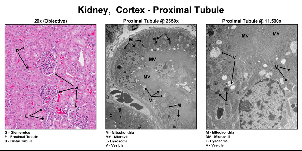
Conventional histopathology uses light microscopy to evaluate stained tissue sections on glass slides, however, some objects are too small to distinguish by light microscopy. 

Electron microscopy (EM) is a technique used in biomedical research for obtaining high resolution images to investigate ultrastructural details.

GLP TEM Lab Features

  • In-house automated processors and grid stainers capable of high throughput epoxy resin processing and staining
  • Resin-embedded sections cut using modern ultramicrotomes
  • Toluidine blue staining performed for selection of specific structures for analysis by EM, or for neurotoxicity analysis when electron microscopy may not be needed
  • High quality Transmission Electron Microscopy (TEM) images for review by in house expert TEM pathologists or electronically transferred for client analysis
  • Good Laboratory Practices (GLP) compliant end-to-end; image capture software has been validated to ensure electronic data regulatory compliance. 

When to Include TEM Work

  • Regulatory advice received e.g. the FDA has alerted to expect changes, such as in the mitochondria, and proactive analysis may speed up the IND process
  • Cationic amphiphilic drug with known history and reason to suspect phospholipidosis
  • Biologics—suspect immune complex deposition e.g. kidney glomeruli
  • Observed abnormal cellular presentation (lesion) e.g. vacuoles, or myelin figures in the neurons of brain tissue
Kidney, Cortex - Proximal Tubule

Let's start a conversation

Contact Us