Flow Cytometry

Contact Us

The most recent breakthroughs in cancer research and treatment have been in the field of immuno-oncology, using novel immunotherapies to bolster the host’s immune system to more effectively target and destroy tumor cells. Among the list of FDA-approved therapeutics with immunoregulatory activity are antibody and cytokine-based immunotherapies, small molecule drugs, and cell-based therapy. The success that these and other therapies have had at prolonging survival has placed emphasis on the development of new single agent and combination therapies with greater efficacy at treating cancer.

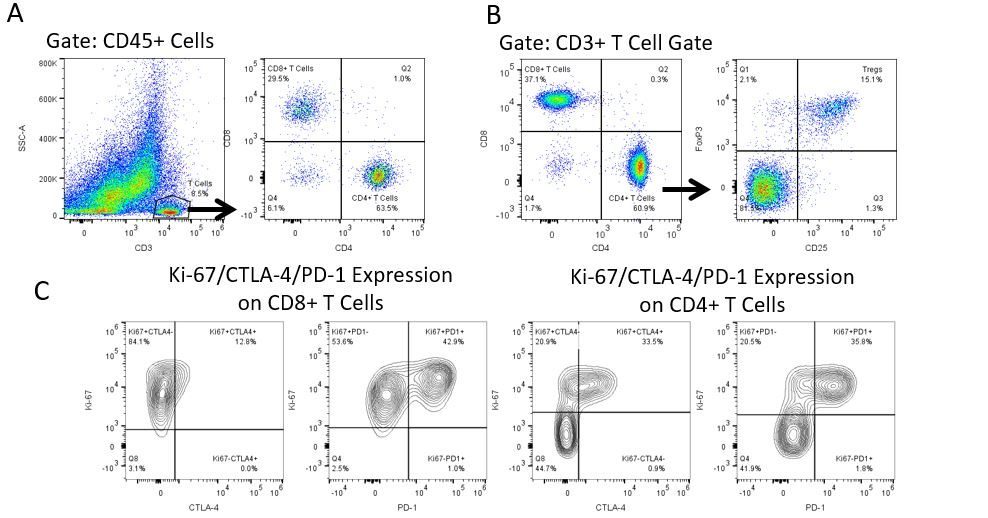
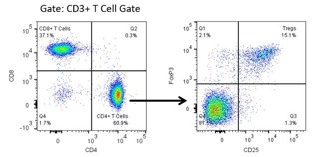
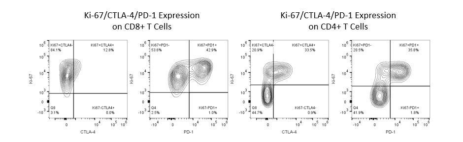
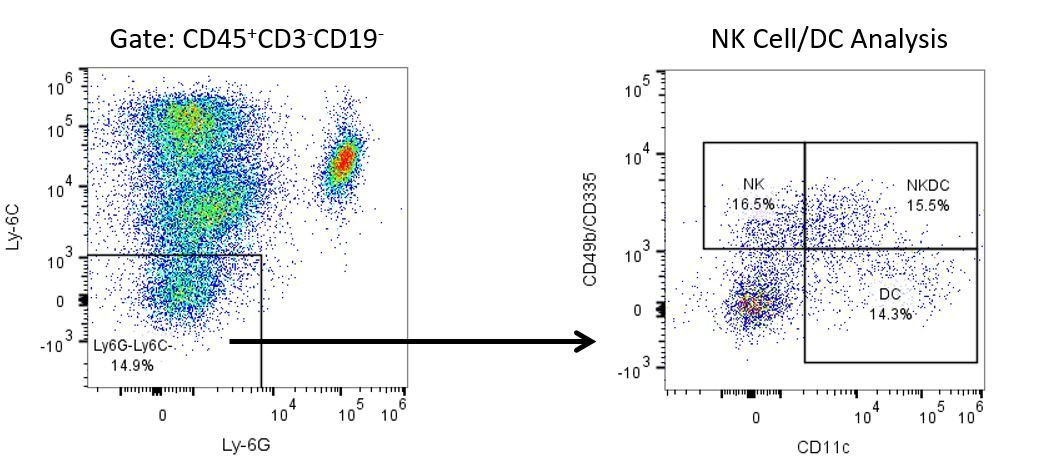
Productive development of immunotherapies requires high-throughput and robust quantitative analysis of the immune system in the tumor micro-environment, peripheral blood, and other host tissues.

To meet this need, our contract flow cytometry service, provides you with an advanced, state-of-the-art analytical flow cytometry resource to support all aspects of your drug development needs. Run your sample generation studies with us or overnight-ship us your preclinical or non-CLIA regulated clinical samples and we’ll take care of the flow cytometry for you.

Basic to Comprehensive Immune Profiling

With over 50 years of combined expertise, our full-service analytical team can add an ex vivo flow cytometry arm to any in vivo study. Our list of standardized antibody panels (see below) have been validated in multiple in vivo cancer models and provide basic to comprehensive analysis of a wide range of lymphoid and myeloid lineage immune subsets to capture shifts that occur in the immune response triggered by in vivo test agent treatment. See a list of our panels below.

  • Basic T Cell Panel

    CD4+, CD8+, and regulatory T cell analysis combined with immune checkpoint/activation marker interrogation using the T Cell Panel

    The Comprehensive T Cell panel combines the utility of the Basic, Regulatory, and T Cell Activation/Exhaustion panels into one comprehensive analysis of the T cell profile within tissues. The data above displays (A) CD4+/CD8+ T cell subset analysis in murine tumors using a 4T1 breast carcinoma model, (B) regulatory T cell analysis in spleens from CT.26 colorectal adenocarcinoma bearing mice, and (C) CTLA-4, PD-1, and Ki-67 expression levels in T cell subsets from A20 (B cell lymphoma) cell line-derived tumors.
  • Basic T & B Cell Panel

    T cell and B cell subset analysis in murine spleen.

    The Basic T & B Cell Panel can be utilized to quantify CD4+ and CD8+ T cells as well as B cells fractions in heterogeneous samples. Only 6 fluorescence channels are occupied by this panel, which leaves several channels available for further cell characterization by dropping in additional antibodies. In the study above, CD3+ T cells and CD19+ B cells were analyzed in the Live Cell gate. T cells were further subdivided into CD4+ and CD8+ populations.

  • Regulatory T Cell Panel

    Analysis of T cell activation in the murine A20 B cell lymphoma model

    One challenge faced during the development of new immunotherapies is overcoming the loss of anti-tumor activity that occurs in T cells within the tumor microenvironment. A state referred to as T cell exhaustion, which can be identified based on the relative expression of different immune checkpoints and activation markers. The T Cell Activation/Exhaustion panel is an expansion of the Basic T Cell panel and can be used to measure expression for select key biomarkers in different tissues. In the study above, expression of immune checkpoint proteins CTLA-4 and PD-1 as well as the surrogate proliferation marker Ki-67 were measured in T cell subsets within A20 cell line-derived tumors.

  • T Cell Activation/Exhaustion Panel

    Analysis of T cell activation in the murine A20 B cell lymphoma model

    One challenge faced during the development of new immunotherapies is overcoming the loss of anti-tumor activity that occurs in T cells within the tumor microenvironment. A state referred to as T cell exhaustion, which can be identified based on the relative expression of different immune checkpoints and activation markers. The T Cell Activation/Exhaustion panel is an expansion of the Basic T Cell panel and can be used to measure expression for select key biomarkers in different tissues. In the study above, expression of immune checkpoint proteins CTLA-4 and PD-1 as well as the surrogate proliferation marker Ki-67 were measured in T cell subsets within A20 cell line-derived tumors.

  • Comprehensive T Cell Panel

    CD4+, CD8+, and regulatory T cell analysis combined with immune checkpoint/activation marker interrogation using the T Cell Panel

    The Comprehensive T Cell panel combines the utility of the Basic, Regulatory, and T Cell Activation/Exhaustion panels into one comprehensive analysis of the T cell profile within tissues. The data above displays (A) CD4+/CD8+ T cell subset analysis in murine tumors using a 4T1 breast carcinoma model, (B) regulatory T cell analysis in spleens from CT.26 colorectal adenocarcinoma bearing mice, and (C) CTLA-4, PD-1, and Ki-67 expression levels in T cell subsets from A20 (B cell lymphoma) cell line-derived tumors.
  • Natural Killer Cell Panel

    Natural Killer (NK) cell and Dendritic Cell (DC) subset analysis in CT.26 colorectal adenocarcinoma model

    NK cell subsets are well known for their anti-tumor activity and ability to control the growth of many tumors. DCs have both pro- and anti-tumor function and are a target for vaccine-based immunotherapies. NKDC (aka IKDC) have been shown to posses functional characteristics of both NK cells and DCs. The Natural Killer Cell panel can be used to identify theses subsets. The study above was performed to quantify percentages of NK cell, DC, and NKDC subsets within tumor-derived Ly6G-Ly6C- immune cells.

  • Myeloid-Derived Suppressor Cell Panel

    Myeloid-Derived Suppressor Cell (MDSC) analysis in a murine CT.26 colorectal oncology model

    MDSC subsets can influence several pro-tumor responses including the suppression of immune function, and are an attractive target for immunotherapy. They are a heterogeneic cell population. The phenotype of which can be shaped by signals in the tumor microenvironment. The data above demonstrates how the MDSC panel can be used for basic identification of granulocytic (G-) and monocytic  (M-) MDSC subsets in CT.26-derived tumors. CD11b+ myeloid cells were first identified in the CD45+ gate after CD3+CD19+ lymphocyte exclusion. G-MDSC and M-MDSC were further differentiated based on expression of Ly-6G and Ly-6C surface receptors.

  • Expanded Myeloid Panel

    Analysis of myeloid-derived suppressor cell (MDSC), natural killer (NK) cell, and dendritic cell (DC) subsets in a murine CT.26 colorectal tumor model.

    The Expanded Myeloid panel is an expansion of the basic MDSC panel to enable analysis of NK and DC subsets in addition to MDSC populations. In the data shown, these subsets were analyzed in CT.26-derived tumors. Monocytic (M-) and Granulocytic (G-) MDSCs express high levels of Ly-6C and Ly-6G protein respectively (Bright) and co-expressed the CD11b marker. NK and DCs were analyzed in the MDSC exclusion gate and further differentiated based on pan-NK markers (CD49b and CD335) vs DC-specific CD11c protein expression. Red and grey peaks represent CD11b stained and unstained cells respectively.

  • Comprehensive Myeloid Panel

    Myeloid Cell Analysis in tumors using the MI Bioresearch Comprehensive Myeloid Panel

    The Comprehensive Myeloid Panel builds on the Basic Myeloid-Derived Suppressor Cell Panel for a more in depth analysis of myeloid cell populations. It also enables the analysis of the immune inhibitory receptor PD-L1 on both tumor and immune cell subsets. The data above demonstrates how the Comprehensive Myeloid Panel can be used to A) measure PD-L1 expression on tumor and immune cells (4T1 breast carcinoma model), B) Quantify macrophage and dendritic cell subsets (CT.26 colorectal tumor model), and C) characterize MDSC subsets for expression of CD115, which is a receptor that has been shown to directly correlate with suppressive activity (CT.26 colorectal tumor model). This panel can also be helpful in the analysis of neutrophils and monocyte populations (not shown). Red and grey peaks represent cells stained for target antigen and unstained cells respectively.

  • M1 and M2 Tumor-Associated Macrophage Panel

    Analysis of classically (M1) and alternatively (M2) activated tumor-associated macrophages in a CT.26 colorectal adenocarcinoma model

    M1 and M2 macrophages are the two major macrophage groups that reside within the tumor microenvironment. They have opposing functions with regard to cancer progression and are therefore targets for new immunotherapies. The study above demonstrates how the M1 and M2 Tumor-Associated Macrophage panel can be used to profile the ratios of these two groups. To this end, MDSC subsets were excluded so that CD11b+/F480+ macrophages could be identified.  This fraction was analyzed for M2 macrophages (CD206+) and M2 macrophages (CD206-MHCII+).

Let's start a conversation

Contact Us