Author: David Draper, Ph.D., Scientist, In Vitro Operations
Date: January 2018
In this article, we will be presenting phospho-protein detection in solid tumors using phospho-flow cytometry and reviewing the analysis of cabozantinib-induced inhibition of PI3K/AKT and JAK-STAT signaling in tumor vs. host immune cells.
The in vivo efficacy of small molecule inhibitors is often assessed by measuring the phosphorylation state of cell signaling proteins within the pathways the drug targets. This can be a challenge when analysis of solid tumors is required. In pre-clinical research that utilizes rodent models, the most common approaches for tumor analysis are immunoblot and ELISA-based techniques. However, these bulk analysis methods have two main drawbacks. Their relative ability to measure multiple targets simultaneously is suboptimal. More importantly, they are unable to analyze targets in multiple distinct subsets within the heterogeneous tumor sample. This can be important when the target signaling protein has opposing functions in different cell types in the tumor microenvironment such as immune cells vs. tumor cells, and even critical if drug-induced effects occur in one but not both subsets.
Labcorp has developed a novel phospho-flow-based platform that simultaneously analyzes multiple cell signaling targets in both the host immune cell and the tumor cell compartments derived from solid tumors. This is accomplished by distinguishing the tumor cells from the infiltrating host immune cells by immunophenotyping for the pan-hematopoietic cell marker CD45, prior to phospho-protein analysis. To demonstrate the utility of this platform, the levels of phosphorylated STAT5 and the downstream target of AKT (S6) were measured in subcutaneous MV-4-11 tumors harvested from mice treated with the FLT3 inhibitor cabozantinib. Figure 1 reveals that cabozantinib reduces the levels of both pS6 and pSTAT5 in the human (h)CD45+ tumor cells. No changes in the levels of these phospho-proteins were observed in the mouse (m)CD45+ host immune cells (not shown). Thus, insight into PI3K/AKT and JAK-STAT signaling activity is gained, which are well documented pathways that regulate both tumor growth and host immunity, and are attractive targets for therapeutic intervention.
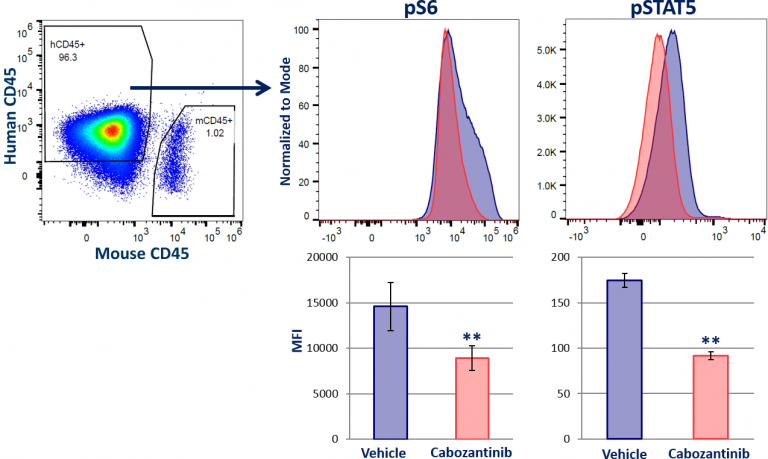
Fig. 1: Solid Tumor Phospho-Flow Analysis of STAT5 and MAPK Signaling.
MV-4-11 is a model for acute myeloid leukemia (AML), a malignancy driven by genetic mutation in one of several genes. The most common event occurs in FLT3 gene, which results in a constitutively active FLT3 protein that drives cancerous cell proliferation by triggering hyper-activation of PI3K/AKT and JAK-STAT signaling.1 Cabozantinib-mediated FLT3 inhibition has been demonstrated to arrest MV-4-11 tumor growth in vivo, which correlated with an inhibition of AKT and STAT signaling in vitro.2 Although overactive PI3K/AKT and JAK-STAT5 signaling has been well documented to drive tumor growth in many models, the activity of these signaling proteins are also required for T cell activation and the generation of memory T cell formation.3,4,5 This dichotomy emphasizes the importance of being able to simultaneously analyze phospho-proteins in both solid tumor-derived immune and tumor cells.
Tumor phospho-flow at Labcorp has potential to advance the cell signaling field. Solid tissue phospho-flow was described in 2010 in lung and peritoneal tumors.6 Since, reports of other successful approaches have been scarce. More recently, an approach termed DISSECT was paired with cytometry and successfully used to measure phospho-proteins in epithelium samples, and later colorectal tumors.7,8 It should be noted that these approaches require fixation after tissue harvest, which can alter the epitopes that are bound by the fluorescent antibodies used for both immunophenotyping and signaling analysis. This can limit the amount of data the techniques provide. Solid tumor phospho-flow at Labcorp does not require fixation, prior to immuno-staining. This facilitates our ability to target distinct populations for analysis. We are working to expand our capabilities. Ongoing efforts are focused on the development of new solid tumor phospho-flow services that will enable the analysis of in vivo treatment induced effects in specific T cell subsets and tumor cells simultaneously.
Contact us to learn more about solid tumor phospho-flow, as well as information on our expanding list of signaling proteins for which we can provide analysis.
References
1Hospital, M. A.; Green, A. S.; Maciel, T. T.; Moura, I. C.; Leung, A. Y.; Bouscary, D.; & Tamburini, J. (2017). FLT3 inhibitors: clinical potential in acute myeloid leukemia. OncoTargets and therapy, 10, 607.
2Lu, J. W.; Wang, A. N.; Liao, H. A.; Chen, C. Y.; Hou, H. A.; Hu, C. Y.; … & Lin, L. I. (2016). Cabozantinib is selectively cytotoxic in acute myeloid leukemia cells with FLT3-internal tandem duplication (FLT3-ITD). Cancer letters, 376(2), 218-225.
3Vara, J. Á. F.; Casado, E.; de Castro, J.; Cejas, P.; Belda-Iniesta, C.; & González-Barón, M. (2004). PI3K/Akt signalling pathway and cancer. Cancer treatment reviews, 30(2), 193-204.
4Rani, A. & Murphy, J. J. (2016). STAT5 in cancer and imCantrell, D. (2002, February).
5Protein kinase B (Akt) regulation and function in T lymphocytes. In Seminars in immunology (Vol. 14, No. 1, pp. 19-26). Academic Press.
6Lin, C. C.; Huang, W. L.; Su, W. P.; Chen, H. H.; Lai, W. W.; Yan, J. J.; & Su, W. C. (2010). Single cell phospho‐specific flow cytometry can detect dynamic changes of phospho‐Stat1 level in lung cancer cells. Cytometry Part A, 77(11), 1008-1019.
7Simmons, A. J.; Banerjee, A.; McKinley, E. T.; Scurrah, C. R.; Herring, C. A.; Gewin, L. S.; … & Irish, J. M. (2015). Cytometry‐based single‐cell analysis of intact epithelial signaling reveals MAPK activation divergent from TNF‐α‐induced apoptosis in vivo. Molecular systems biology, 11(10), 835.
8Simmons, A. J.; Scurrah, C. R.; McKinley, E. T.; Herring, C. A.; Irish, J. M.; Washington, M. K.; … & Lau, K. S. (2016). Impaired coordination between signaling pathways is revealed in human colorectal cancer using single-cell mass cytometry of archival tissue blocks. Science signaling, 9(449), rs11.
Note: Please note that all animal care and use was conducted according to animal welfare regulations in an AAALAC-accredited facility with IACUC protocol review and approval.
Connect
Let's start a conversation
Contact Us
Labcorp is a leading global life sciences company that includes contract research and developmental services to the pharmaceutical, medical technology, crop protection and chemical industries.