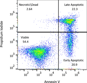
Apoptosis is a normal physiologic process, the program of which is characterized by certain morphologic features, including loss of plasma membrane asymmetry and attachment, condensation of the cytoplasm and nucleus, and internucleosomal cleavage of DNA. FITC Annexin V staining precedes the loss of membrane integrity which accompanies the latest stages of cell death resulting from either apoptotic or necrotic processes. Therefore, staining with FITC Annexin V is typically used in conjunction with a vital dye such as propidium iodide (PI) to identify early apoptotic cells (PI negative, FITC Annexin V positive). Viable cells with intact membranes exclude PI, whereas the membranes of dead and damaged cells are permeable to PI. Let us determine if treatment with your compound includes apoptosis in your samples and receive data regarding the percentage of cells in each sample that are alive, apoptotic, and dead. Our flow cytometry team can perform apoptosis assays on multiple types of samples such as cultured cells, blood, or tissues, and, if desired, analyze specific subsets of cells within each sample.

-
Apoptosis via flow cytometry
-
Cell cyle via flow cytometry
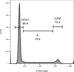
When staining the DNA of a cell with a fluorescent DNA binding agent, such as propidium iodide (PI), the relative amount of DNA in the cell can be measured via flow cytometry and therefore, the stage of the cell cycle can be elucidated. During G0 or G1, a cell contains 46 chromatids; during the S phase, the chromosomes duplicate, and by the end of S phase, G2, or M phase, there are 92 chromatids. The effects of cell cycle checkpoint inhibitors can be confirmed using this method. Our flow cytometry experts are able to confirm the effects of cell cycle checkpoint inhibitors with the method mentioned above, as well as determine the percentage of cells in different stages of the cell cycle in samples such as cultured cells, blood, and tissues.
-
Intracellular signaling via phospho-flow cytometry
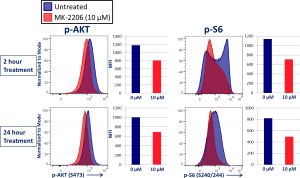
We also offer phospho-flow cytometry, which is a valuable tool for screening new drugs for their effects on the activation of cell signaling pathways in vitro; as well as having the ability to measure drug effects in vivo by multiplexing the technique with immunophenotyping cell surface markers to distinguish analysis in different cell subsets. This platform uses fluorescence-labeled antibodies that recognize proteins only when phosphorylated on specific amino acid residues that regulate their function. Phospho-flow data is highly consistent and reproducible, which in turn creates a unique platform for cell signaling analysis.
Connect
Let's start a conversation
Contact Us
Labcorp is a leading global life sciences company that includes contract research and developmental services to the pharmaceutical, medical technology, crop protection and chemical industries.